Try the questions in the test above first, then open each one here to see the correct answer and why every other option is wrong.
01A hospital is sued for an error made by an employed technologist during routine duties. Under 'respondeat superior', which statement is correct?
Practice question Unit: Professional Practice
ALiability passes entirely to the physician who referred the patient
The referring physician ordered the exam but did not employ or direct the technologist in performing it, so liability does not shift to them automatically.
BOnly the technologist can be held liable, as the person who acted
This ignores the doctrine itself: respondeat superior exists precisely so that the employer can also be held liable for an employee's acts within the scope of employment.
CThe employer can be liable; the technologist stays accountable ✓ Correct answer
Correct. Under respondeat superior the employer can be held vicariously liable for negligence committed during routine duties, but the technologist remains personally accountable for their own professional conduct and can also be named.
DNo one is liable if the technologist was following a verbal order
A verbal order does not excuse a negligent act. The technologist must still perform the task competently and question an unsafe instruction.
Reference: Adler & Carlton, Introduction to Radiologic and Imaging Sciences and Patient Care, 6th ed. (Elsevier), Medicolegal considerations: respondeat superior and personal liability
Ask a friend
02A patient is booked for an elective diagnostic angiogram. Who is responsible for obtaining the patient's informed consent?
Practice question Unit: Professional Practice
AThe nurse who admitted the patient to the ward
The admitting nurse can check that a signed form is present, but cannot explain the specific risks, benefits and alternatives of an angiogram.
BThe physician who will perform the procedure ✓ Correct answer
Correct. Informed consent must be obtained by the physician performing the invasive procedure, because that person can explain its nature, risks, benefits and alternatives and answer questions. Other staff may only witness the signature.
CThe referring physician who ordered the study
The referring physician decides the study is indicated, but does not perform it. Consent for the procedure itself is obtained by the operator who will carry it out.
DThe technologist preparing the angiography room
The technologist may witness the patient's signature, but obtaining consent for an invasive procedure is not within the technologist's role.
Reference: Adler & Carlton, Introduction to Radiologic and Imaging Sciences and Patient Care, 6th ed. (Elsevier), Informed consent: who obtains consent; cf. Saudi Law of Practicing Healthcare Professions (Royal Decree M/59, 1426H), patient consent provisions
Ask a friend
03After a contrast injection, a patient becomes pale and sweaty with BP 80/50 mmHg and a pulse of 45/min, with no rash or wheeze. Which is the most likely reaction?
Practice question Unit: Health and Safety
AContrast-induced nephropathy
Contrast-induced nephropathy is a delayed rise in creatinine over 24–72 hours. It does not cause sudden hypotension and bradycardia minutes after injection.
BHypoglycemic episode
Hypoglycemia can cause pallor and sweating, but it typically produces a fast pulse and is not triggered by contrast. Bradycardia with hypotension points elsewhere.
CVasovagal reaction ✓ Correct answer
Correct. The ACR Manual on Contrast Media distinguishes a vasovagal reaction by hypotension with bradycardia (often pallor, sweating, nausea). Treatment is to elevate the legs, give IV fluids and, if bradycardia persists, atropine.
DAnaphylactic shock
Anaphylactic hypotension is usually accompanied by tachycardia and often by urticaria, wheeze or airway swelling. None of these is present here.
Reference: ACR Manual on Contrast Media (ACR Committee on Drugs and Contrast Media, 2024), Treatment of acute reactions: vasovagal reaction vs anaphylactoid hypotension; https://www.acr.org/Clinical-Resources/Clinical-Tools-and-Reference/Contrast-Manual
Ask a friend
04A woman who had an abdominopelvic CT is later found to have been 9 weeks pregnant. The medical physicist estimates the fetal dose at 25 mGy, and she asks whether the radiation is a reason to end the pregnancy. ICRP Publication 84 states that termination is NOT justified on radiation-risk grounds below what fetal dose?
Practice question Unit: Health and Safety
A1 mGy
1 mGy is in the range of the public dose limit and many low-dose exams. It is far below any level at which termination is even discussed.
B10 mGy
10 mGy is about the fetal dose of a single pelvic CT. Exceeding it calls for a dose estimate and counselling, not for considering termination.
C100 mGy ✓ Correct answer
Correct. ICRP 84 states that termination of pregnancy at fetal doses below 100 mGy is not justified on the basis of radiation risk. At 25 mGy she can be reassured that the radiation risk does not warrant ending the pregnancy.
D1000 mGy
1000 mGy is far too high a threshold. Malformation and neurodevelopmental risks rise well below 1 Gy, so ICRP sets the decision point an order of magnitude lower.
Reference: ICRP Publication 84 (2000), Pregnancy and Medical Radiation, Ann. ICRP 30(1), summary point on termination below 100 mGy; https://www.icrp.org/publication.asp?id=ICRP%20Publication%2084
Ask a friend
05A radiographer declares her pregnancy to her employer. Under ICRP 103 and IAEA GSR Part 3, what dose restriction applies to the embryo or fetus for the remainder of the pregnancy?
Practice question Unit: Health and Safety
A20 mSv per year
20 mSv per year (averaged over 5 years) is the occupational effective-dose limit for the worker herself, not the protection level for the embryo or fetus.
B50 mSv in a single year
50 mSv is the maximum occupational dose in any single year within the 5-year average. Applying it to the fetus would give no extra protection after declaration.
CAbout 1 mSv to the fetus ✓ Correct answer
Correct. ICRP 103 and IAEA GSR Part 3 require that, once pregnancy is declared, the embryo or fetus receive broadly the same protection as a member of the public, i.e. not more than about 1 mSv for the remainder of the pregnancy.
D0.5 mSv per month to the fetus
0.5 mSv per month is the US NCRP monthly guidance for the embryo/fetus, not the ICRP/IAEA restriction named in the question.
Reference: ICRP Publication 103 (2007), §§185–186 (pregnant workers: about 1 mSv to the embryo/fetus after declaration); IAEA GSR Part 3 (2014), paras 3.113–3.114; https://www.icrp.org/publication.asp?id=ICRP%20Publication%20103
Ask a friend
06A grid has lead strips 2.4 mm high separated by 0.3 mm interspaces. What is its grid ratio?
Practice question Unit: Operation of Equipment
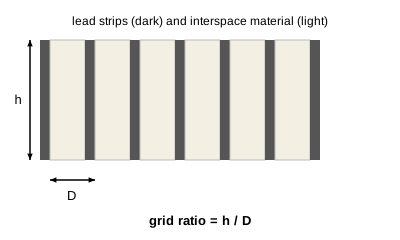
A8:1 ✓ Correct answer
Correct. Grid ratio = height of the lead strips ÷ width of the interspace (h/D) = 2.4 mm ÷ 0.3 mm = 8, i.e. 8:1.
B2.1:1
2.1 comes from subtracting the interspace from the strip height (2.4 − 0.3). Grid ratio is a quotient, not a difference.
C12:1
A 12:1 grid with 0.3 mm interspaces would need strips 3.6 mm high (12 × 0.3). These strips are only 2.4 mm.
D2.7:1
2.7 comes from adding the two values (2.4 + 0.3) instead of dividing height by interspace width.
Reference: Bushong, Radiologic Science for Technologists: Physics, Biology, and Protection, 12th ed. (Elsevier, 2021), Ch. Control of scatter radiation: grid ratio = h/D
07A knee is taken at 70 kVp and 20 mAs. Using the 15% rule, which new technique keeps receptor exposure about the same?
Practice question Unit: Operation of Equipment
A80 kVp and 10 mAs ✓ Correct answer
Correct. Raising kVp by 15% (70 × 1.15 ≈ 80 kVp) roughly doubles receptor exposure, so mAs is halved (20 → 10 mAs) to keep exposure about the same.
B60 kVp and 10 mAs
Lowering kVp by about 15% already halves exposure; halving mAs as well cuts it to about one quarter, so the image would be underexposed.
C85 kVp and 20 mAs
85 kVp is about a 21% increase with unchanged mAs, so exposure rises well above double. mAs must be reduced when kVp is raised.
D80 kVp and 40 mAs
A 15% kVp rise doubles exposure and doubling mAs doubles it again, giving about four times the original receptor exposure.
Reference: Carlton & Adler, Principles of Radiographic Imaging: An Art and a Science, 6th ed. (Cengage), Ch. Kilovoltage: the 15 percent rule; Bushong, Radiologic Science for Technologists, 12th ed.
Ask a friend
08A 26-year-old woman is referred with a new palpable lump in her left breast. According to the ACR Appropriateness Criteria, what is the usual first imaging test?
Practice question Unit: Protocol Management
ATargeted breast ultrasound ✓ Correct answer
Correct. For a palpable mass in a woman under 30, the ACR Appropriateness Criteria rate targeted ultrasound as usually appropriate as the first test: young breasts are dense, ultrasound reliably separates cysts from solid masses, and it uses no ionizing radiation.
BContrast-enhanced breast MRI
Breast MRI is a problem-solving or high-risk screening tool. It is rated usually not appropriate as the initial test for a palpable lump.
CTc-99m sestamibi molecular breast imaging
Molecular breast imaging is a supplemental nuclear-medicine test with a whole-body radiation dose. It has no role as the first test for a palpable lump.
DDiagnostic mammography
Mammography is the usual first test from age 40 and is weighed with ultrasound at 30–39. Under 30, dense tissue lowers its sensitivity, so it is reserved for when ultrasound findings are suspicious.
Reference: ACR Appropriateness Criteria: Palpable Breast Masses (2022 revision), variant: women younger than 30 years; https://www.acr.org/Clinical-Resources/Clinical-Tools-and-Reference/Appropriateness-Criteria
Ask a friend
09A contrast-enhanced CT request states 'no allergies', but on questioning the patient reports a severe reaction to iodinated contrast during a previous scan. What should the technologist do?
Practice question Unit: Protocol Management
AHalve the contrast dose and go ahead with the scan
Allergic-like contrast reactions are not dose-dependent, so halving the dose does not make a repeat reaction meaningfully less likely.
BScan without contrast instead
Dropping contrast changes the examination and may make it non-diagnostic. That protocol decision belongs to the radiologist, not to the technologist acting alone.
CInject as requested on the form
The patient's own history overrides the form. Injecting despite a reported prior severe reaction exposes the patient to a known, avoidable risk.
DHold the injection; tell the radiologist ✓ Correct answer
Correct. The ACR Manual on Contrast Media lists a prior allergic-like reaction to the same class of contrast as the strongest risk factor. The technologist withholds the injection and informs the radiologist, who decides on an alternative exam, a different agent and/or premedication.
Reference: ACR Manual on Contrast Media (ACR Committee on Drugs and Contrast Media, 2024), Patient selection and preparation: prior allergic-like reaction; premedication; https://www.acr.org/Clinical-Resources/Clinical-Tools-and-Reference/Contrast-Manual
Ask a friend
10A small left pneumothorax is suspected, but the patient cannot stand or sit erect. Which decubitus projection, with a horizontal beam, best demonstrates it?
Practice question Unit: Protocol Management
ARight lateral decubitus ✓ Correct answer
Correct. Free air rises to the highest point, so the affected left side must be up: the patient lies on the right side (right lateral decubitus) and air collects along the left lateral chest wall, where it is seen with a horizontal beam.
BProne, with a horizontal beam
Prone positioning sends air to the posterior chest, where it overlaps the lung and spine on a frontal view and is hard to see.
CLeft lateral decubitus
Left lateral decubitus puts the affected side down, so the air moves away from the left wall. This position is used to show fluid on the left, not air.
DSupine, with a vertical beam
On a supine film taken with a vertical beam, air collects anteriorly and a small pneumothorax is easily missed (it may show only as a deep sulcus sign).
Reference: Lampignano & Kendrick, Bontrager's Textbook of Radiographic Positioning and Related Anatomy, 10th ed. (Elsevier), Chest: lateral decubitus position (AP projection) for air and fluid levels
Ask a friend

